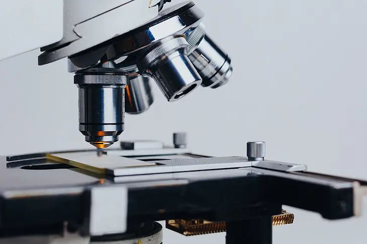
Optical microscopy is an observation technique capable of producing magnified images of objects or their details that are too small to be observed with the naked eye. The instrument used to perform this technique is called an optical microscope. To see an object clearly, a sharp image must be formed on the retina of the human eye.
The lens has the properties of a converging lens that can change its shape and therefore its focal distance; its elasticity allows the eye to adjust for distinct vision at distances ranging from a minimum of about 250 mm, called the near point, to infinity, called the remote point.
When one wants to examine a very small object in detail, one brings it as close as possible to the eye so that the viewing angle is as small as possible and the image on the retina is as large as possible. The smallest distance to which the eye can adapt for clear vision, however, is that of the near point. To overcome this limitation, one resorts to the use of a complex system of lenses called a microscope.
Optical Microscopy
In optical microscopy, lasers are used as light sources to enhance image clarity, resolution, and contrast, especially in advanced imaging techniques. The most commonly used types of lasers in optical microscopy include diode lasers, argon-ion lasers, He-Ne lasers, and femtosecond lasers, each selected based on the imaging requirements and the type of microscopy being performed.
Diode lasers are widely used in fluorescence and confocal microscopy due to their compact size, energy efficiency, and availability in a range of wavelengths. They are ideal for exciting specific fluorophores and are commonly used in live-cell imaging, laser scanning microscopy, and routine fluorescence applications.
Argon-ion lasers emit light at specific visible wavelengths (commonly 488 nm and 514 nm) and are well-suited for exciting popular fluorescent dyes. These lasers are often used in confocal and multi-photon microscopy for high-resolution imaging, particularly in biological samples.
He-Ne (Helium-Neon) lasers, typically emitting at 543 nm or 633 nm, are valued for their excellent beam stability and coherence. They are used in interference-based microscopy techniques such as interferometric or phase-contrast imaging, and also for exciting certain red fluorophores in fluorescence microscopy.
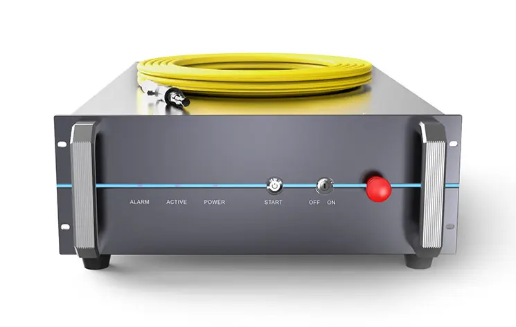
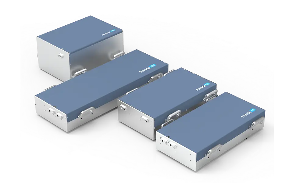
Femtosecond lasers, which emit ultrashort pulses, are essential in multiphoton microscopy. These lasers allow deep tissue imaging with minimal photodamage and are used in neuroscience, developmental biology, and other fields requiring high-resolution, 3D imaging of thick samples.
The type of laser used in optical microscopy depends on the specific technique, the sample type, and the resolution needed. Diode and argon-ion lasers are common for fluorescence-based imaging, He-Ne lasers are preferred for stability and interferometry, and femtosecond lasers are used for advanced, high-depth imaging methods.
In optical microscopy, lasers are used as light sources to enhance image clarity, resolution, and contrast, especially in advanced imaging techniques. The most commonly used types of lasers in optical microscopy include diode lasers, argon-ion lasers, He-Ne lasers, and femtosecond lasers, each selected based on the imaging requirements and the type of microscopy being performed.
Diode lasers are widely used in fluorescence and confocal microscopy due to their compact size, energy efficiency, and availability in a range of wavelengths. They are ideal for exciting specific fluorophores and are commonly used in live-cell imaging, laser scanning microscopy, and routine fluorescence applications.
Argon-ion lasers emit light at specific visible wavelengths (commonly 488 nm and 514 nm) and are well-suited for exciting popular fluorescent dyes. These lasers are often used in confocal and multi-photon microscopy for high-resolution imaging, particularly in biological samples.
He-Ne (Helium-Neon) lasers, typically emitting at 543 nm or 633 nm, are valued for their excellent beam stability and coherence. They are used in interference-based microscopy techniques such as interferometric or phase-contrast imaging, and also for exciting certain red fluorophores in fluorescence microscopy.
Femtosecond lasers, which emit ultrashort pulses, are essential in multiphoton microscopy. These lasers allow deep tissue imaging with minimal photodamage and are used in neuroscience, developmental biology, and other fields requiring high-resolution, 3D imaging of thick samples.
The type of laser used in optical microscopy depends on the specific technique, the sample type, and the resolution needed. Diode and argon-ion lasers are common for fluorescence-based imaging, He-Ne lasers are preferred for stability and interferometry, and femtosecond lasers are used for advanced, high-depth imaging methods.